
NORMAL EEG WAVES SOFTWARE
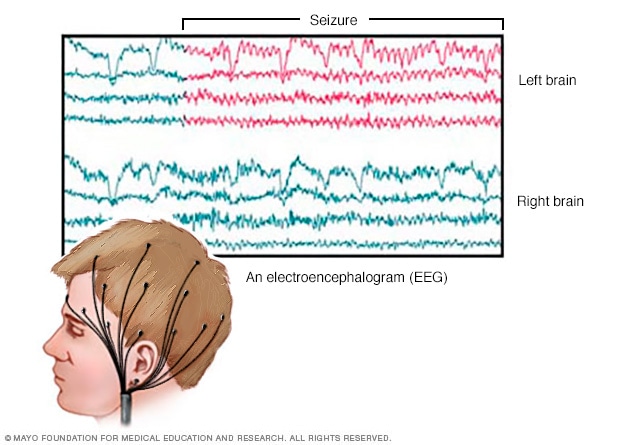
Note that "how big" each second appears differs from one example to the next this is because they were collected on different size screens, and software screen calibration affects the appearance similar to how page speed does (although they are two separate entities) On these tracings, each dark gray line is one second, with the lighter grey lines in between being 1/5 of a second each try counting how many of each wave are seen per second to confirm the frequency on each example. Below are a set of examples of one predominant frequency per page, but note the multiple intermixed frequencies on each. Instead, you'll see a mixture of frequencies overlying one another for example, you can have beta activity on top slower delta activity, which could be termed delta with overriding beta, or delta with admixed beta. Realistically, frequencies on EEG will almost never be as clean as the above examples. Of note, there is technically an even faster frequency, gamma (30 Hz and above) but this is not seen with physiologic activity on scalp EEG. More diffuse beta activity can be found most often with benzodiazepine use. Beta is 13-30 Hz, and is most commonly seen in the setting of myogenic (muscle) artifact in the frontal regions. It is the hallmark frequency of the normal awake adult brain, to the point that the posterior dominant rhythm (PDR), a key finding of the normal background, used to be called the alpha rhythm. However, theta re-emerges often in drowsy periods, and is the hallmark of some normal findings including rhythmic temporal theta of drowsiness (RMTD).Īlpha is 8-13 Hz, and is perhaps the frequency you'll come to know and love best. Theta ranges from 4-8 Hz, and tends to be more prominent in childhood than adulthood. It is the de facto finding of slow wave sleep, where it's seen diffusely, but is also commonly found overlying structural abnormalities such as tumors, or over the bifrontal regions in some patients with encephalopathy. Delta is the slowest at 0-4 Hz, and generally speaking should not be present in a normal awake brain. There are four main frequencies of the human brain seen on scalp EEG, in increasing order: delta, theta, alpha and beta. Frequency describes how many waves there are per second, and is measured in hertz (Hz). Recognizing the frequency of the waveforms is fundamental to interpreting EEG.


 0 kommentar(er)
